Cortical variability
Jump to navigation
Jump to search


(CC) Image: Lefèvre and Mangin, 2010
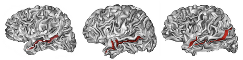
Cortical folding pattern of the human brain, highlighting the anatomical variability of the superior temporal sulcus, which may consist of one (left), two (middle) or three (right) parts. Note that other cortical structures also exhibit considerable variation.
Cortical folding pattern of the human brain, highlighting the anatomical variability of the superior temporal sulcus, which may consist of one (left), two (middle) or three (right) parts. Note that other cortical structures also exhibit considerable variation.
Cortical variability describes the anatomical variability of the cerebral cortex. It manifests itself in variations of the size, shape or number of cortical structures, e.g. the number or shape of sulci and gyri, and is typically expressed in terms of the statistical variation of grey matter volume, cortical thickness, gyrification, cortical surface area, cortical connectivity or related brain morphometric measures across a set of brains or brain images.
The term is usually used in reference to normal healthy adults but it can also refer to other groups of subjects, including patients with brain disorders. Besides the health state, cortical variability is a function of multiple factors, including age, gender, ethnicity and species.