Snake venom
{{dambigbox}Snake venom|Snake}}
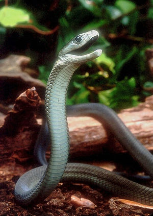
Snake venom is a highly modified saliva that contains many different powerful toxins. There are at least 2.500 species of snakes living at the present time of which over 600 are known to produce venom. Unlike most other predators, all snakes swallow prey whole, so are especially vulnerable to injury if their prey animals are active. Most snake venoms contain specific proteins that (1) paralyze the prey so that it no longer moves (2) interfere with normal blood clotting mechanisms so that the animal goes into shock and (3) begin the process of digestion by breaking down the tissues of the prey animal. Venom also helps deter predators, and is an important defense mechanism for the snake.
Introduction
The process of introducing venom into a victim is called "envenoming". Envenoming by snakes is most often through their bite, but some species, like the 'spitting cobra', use additional methods such as squirting venom onto the mucous membranes of prey animals (eyes, nose, and mouth). Venoms differ from snake species to snake species, and seem to be specialized to dispatch the particular kinds of animals that make up that snake's preferred diet. The great majority of the many biological toxins in snake venom are proteins: some haveenzymatic activity, some can block nerve or muscle cell receptors, and some have activity in the protein cascades for coagulation, complement fixation or inflammation. Effects of snake venom in the tissues envenomated by the bite are called local effects. Other actions arise from toxins transported through the blood vessels or through lymph vessels, and are called systemic effects.
With advances in molecular biology, a general schema for the expression of such proteins by genes in the specialized salivary gland cells that secrete venom has become apparent. The components of the venom may even change over the course of a snake's life, in species (for example, certain rattlesnakes of the genus Crotalus) that rely on one set of prey animals as juveniles (cold-blooded lizards and other small exotherms), and a different set of prey animals as adults (warm-blooded rodents).[1]
Toxicity: LD50
Toxicity of venoms is usually expressed by the LD50: the lowest dose that kills 50% of a group of experimental animals (most often rodents). That dose varies not just between the venoms tested, but also depends on which species of prey animals receive the venom. Generally, the most toxic venom is the one with the lowest LD50. However, some snakes have venoms that are quite specialized for certain types of prey. Few studies have used the natural prey of a snake species, which would involve capturing a number of wild animals. Instead, most research has used inbred strains of laboratory animals. Human susceptibility to a snake venom is generally estimated from the LD50 for rodents. The next factor in assessing the danger of a particular species of snake is the dose of venom that is actually introduced into the tissues. Some types of snakes have an extremely efficient mechanism of injecting venom with a single strike, others have poor success in doing so. The amount of venom produced by snakes that is available for secretion with a bite also varies between kinds of snakes, and between individuals (usually by size) of any one species.
Venom characteristics and delivery of venom according to snake family
The venomous snakes are represented in only four families. There are variations in the methods of envenomation according to family.
Atractaspididae (atractaspidids)
(common names of well-known members: burrowing asps, mole vipers, 'stilleto snakes')
Colubridae (colubrids)
(common names of well-known members: boomslang)
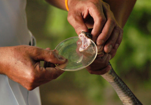
This family of snakes contains about 2/3 of all living species. A minority have somewhat enlarged grooved teeth at the back of the upper jaw for delivering venom under low pressure. This unsophisticated system for venom delivery makes it more difficult for scientists to collect colubrid venom for chemical studies than the venom from vipers and most elapids, which inject venom through front fangs under higher pressure. Often, couloubrid venoms were collected only in relatively small quantities and with impurities from other mouth contents from the snake. As more recent collection methods have been devised that overcome some of these problems, researchers have discovered that earlier assumptions about the venom contents were sometimes mistaken. For example, Phospholipase A2 (PLA2),which had been thought to be lacking in venoms in this family has now been detected in at least two species
"Some venoms show high toxicity toward mice, and others are toxic to birds and/or frogs only. Because many colubrids feed on non-mammalian prey, lethal toxicity toward mice is probably only relevant as a measure of risk posed to humans. At least five species (Dispholidus typus, Thelotornis capensis, Rhabdophis tigrinus, Philodryas olfersii and Tachymenis peruviana) have caused human fatalities."[2]
Elapidae (elapids)
(common names of well-known members: cobras , kraits, coral snakes, mambas, sea snakes, sea kraits, Australian elapids)
The venom of elapid snakes is notorious for the potency of its neurotoxins. These snakes have similarities in their XXXXX. Venomous elapid snakes greatly range in size, aggressiveness, and in habitat. "The king cobra (Ophiophagus hannah) is the world’s longest venomous snake, growing up to 5.5 m (18.5 ft). The main constituent of king cobra venom is a postsynaptic neurotoxin, and a single bite can deliver up to 400–500 mg of venom, ...about fifteen thousand times the LD50 dose for mice. The world’s most venomous snake is the Australian elapid small-scaled snake (Oxyuranus microlepidotus), can deliver up to 100 mg of venom with an LD50 for mice of 0.01 mg.kg)1, giving up to 500 000 LD50 mice doses. [3].
Although sea snakes have some of the world's most potent venom, the numbers of human fatalities from snake bites is apparently limited by their marine environment and behavior (more coming with references).
For prey animals, and in cases of defensive behavior towards humans, "neuromuscular paralysis usually occurs with elapid (cobra, krait, and mamba) envenomation." [4], however, many elapid snakes have venoms that also include toxins that cause bleeding. For example, the venom of , all contain metalloproteinases that interfere with platelet aggregation.
Besides neurotoxins and metalloproteinases, there are additional types of bioactive proteins and polypeptides that are common in elapid venom. "A second group of toxins are cell membrane poisons that act in a general fashion, but their chief effect is on the heart, producing arrhythmias and impaired contractility. The third group of toxins contains enzymes that break down protein and connective tissue. These necrosis-producing toxins are typical of the venom from the spitting cobras (Naja spp.) of Africa, China, and Sumatra." [5]
Viperidae (viperids)
(common names of well-known members: pitless vipers, pit vipers)
Bites by snakes of the family Viperidae often induce local breakdown of muscle and tissues which may result in permanent deformity in the region of the bite (Myotoxic phospholipases).[6] Some types of vipers inject venom that travels though the bloodstream and breaks down muscle cells systemically, with relatively little reaction at the site of the bite, but enough muscle cells throughout the body release their contents into the victim's bloodstream to cause a condition known as rhabdomyolysis. In rhabdomyolysis (literally rhabdo=rod , myo=muscle cell, lysis= breaks apart) the large iron-containing protein myoglobin is released into the circulation (myoglobulinemia). When myoglobin reaches the kidney, the renal system attempts to filter it out of the blood. If the amount of myoglobin is very large, acute renal failure results, and the blood is no longer properly filtered of even normal body wastes by the kidneys.
The common names of vipers frequently fail to identify an actual species. For example, the name, Rock viper refers to two entirely different kinds of snakes.
Crotalinae (crotalines)
(common names of well-known members: pit vipers, including lanceheads, moccasins, rattlesnakes)
Pit viper venom characteristically contains a potent mix of enzymes that produce an emphatic degree of tissue destruction at the site of the bite. As with most venoms, there can be both local and systemic effects. However, unless a bite by a pit viper is "dry" (meaning no venom injected), there will ordinarily be marked inflammation at the site of the bite and possibly systemic effects.
Rattlesnakes range in size from small (pigmy rattlesnakes, Sistrurus) to large (many species of Crotalus, such as the Eastern diamondback, Crotalus adamanteus). Most pit vipers are potentially very active and aggressive snakes. The strike can be lightning quick, measured in one study as under 50ms [7].
Effects of Venom
Snake venoms contain molecules that are biologically active. The poison gland of snakes adds these molecules to saliva, which is the digestive juice produced by the mouth of most all land vertebrates. In nonvenomous snakes, and in other creatures, such as we humans, saliva moistens the food and initiates digestion with enzymes. In venomous snakes, the toxic molecules added to their specialized venom include some of the most powerful substances known in their effects on biological systems.
Some toxins take effect at the site of the bite, others are only active in certain tissues and cause their harmful effects once they reach these tissues through the blood stream. The effects of the snake venom on that victim are not always direct, sometimes the toxic substances trigger cascades of reactions, that, like a toppling row of dominoes, lead to many cumulative disruptions.
Shock
- Hemorrhage and intravascular coagulation: disruption of the normal blood clotting pathways
Many components in snake venom disrupt normal blood flow and normal blood clotting (coagulation). Some common enzymes in snake venoms increase bleeding by preventing the formation of clots, and others by breaking down established clots. Both of these types of enzymes include metalloproteases. Other toxins increase 'bleeding time' by inhibiting the aggregation of platelets, the small odd-shaped blood cells that collect at the site of a tear in a blood vessel and form a plug to close it. Profound loss of blood can cause hemorrhagic shock, and disable a prey animal. When many tiny blood clots form in the bloodstream there is a pathological condition known as disseminated intravascular coagulation (DIC), which also causes shock. Some enzymes in snake venom set off DIC in the bloodstream of their envenomated prey by interfering with the activity of serine proteases involved in the regulation of hemostasis.
- Infarction: Stroke and Heart Attack
Toxins that set off clotting within the blood vessels of envenomated animals can cause both stroke and heart attacks. Infarction is a medical term that means death to tissues because of a block in their blood supply, and clots within the arteries of the neck and brain, as well as the coronary arteries can deprive the blood supply enough to cause infarctions in these organs.
Paralysis
Some proteins secreted in snake venoms are toxins that affect nerves (neurotoxins) and the contractibilty of muscle. Most neurotoxins in snake venoms are too large to cross the blood-brain barrier, and so they usually exert their effects on the peripheral nervous system rather than directly on the brain and spinal cord. Many of these neurotoxins cause paralysis by blocking the neuromuscular junction. In fact, biologists first learned some of the details of how the neuromuscular junction normally functions by using purified snake venoms in physiology experiments.
The Neuromuscular Junction
The neuromuscular junction is the microscopic connection between a motor nerve fiber and a muscle fiber, and is a type of synapse. Muscle contractions are normally regulated by the electrical activity of large nerve cells in the spinal cord and brainstem, called motor neurons or motoneurons. These neurons have long axons ('nerve fibres') that end in contact usually with just a single muscle fibre. The axon endings make a specialized contact with the muscle fiber, that is very like the synapses 'synaptic contacts' between nerve cells in the brain. This contact zone is called the neuromuscular junction, and on both the muscle side and the nerve side of this junction there are specialized structures and specific proteins for regulating the passage of information from neuron to muscle fiber. The nerve ending is filled with small synaptic vesicles that contain neurotransmitters - chemical messengers. When the brain gives the command to move a muscle, electrical signals (action potentials) are propagated down the motoneuron axons to the endings. The endings are depolarized by these signals, and as a result voltage-sensitive calcium channels open in the nerve ending. This calcium entry causes some of the synaptic vesicles to fuse with the nerve cell membrane, causing them to release their chemical contents into the narrow cleft between nerve ending and muscle fiber. The most important of these messengers at the neuromuscular junction is acetylcholine. Across this tiny space between the nerve ending and the muscle cell, the acetylcholine molecules bind to other molecules - the acetylcholine receptor molecules (specifically, muscle-type nicotinic acetylcholine receptors). This receptor is a ligand-gated ion channel; when acetylcholine binds to it, the channel opens, allowing sodium to enter the muscle cell. The inflow of sodium ions causes the muscle fiber to become depolarized and as a result, voltage-sensitive calcium channels open, allowing calcium to enter. As calcium enters, it triggers further calcium release from stores inside the cell (in the sarcoplasmic reticulum), resulting in a 'wave' of calcium that spreads throughout the muscle fiber. The calcium interacts with filaments inside the muscle cell called myofibrils, causing them, and as a result the whole muscle fiber, to contract.
The effect of acetylcholine is normally very short lived, as it is rapidly destroyed by acetylcholinesterase, an enzyme produced both by the muscle fibres and by the motoneurons that very efficiently breaks down the acetylcholine. Without acetylcholinesterase, enough aceytlcholine would remain in the cleft between nerve fiber and muscle cell to keep reactivating the muscle contraction mechanism for a long time, producing a form of tetany.
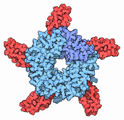
Neurotoxins in snake venom can block transmission of acetylcholine from nerve to muscle at the side of the nerve ending (pre-synaptic literally, before the synapse), or affect the activity of the muscle fiber past the synapse (post-synaptic literally after the synapse). Most commonly, the postsynaptic method of producing paralysis is an anti-cholinesterase toxin in venom that prevents acetylcholinesterase from degrading the acetylcholine. Most snake venoms contain toxins that cause paralysis by both methods: pre and postsynaptic interference. [8]. Presynaptic neurotoxins are commonly called ß-neurotoxins and have been isolated from venoms of snakes of families Elapidae and Viperidae. ß-Bungarotoxin was the first presynaptically active toxin to be isolated from Bungarus multicinctus (banded krait), which is an elapid. ß-bungarotoxin has a phospholipase subunit and a K+ channel binding subunit, and their combined effects are to destroy sensory and motor neurons [9] The banded krait venom also contains alpha-bungarotoxin, which binds to nicotinic acetylcholine receptors, thus preventing acetylcholine from doing so (i.e. it is a receptor antagonist), and kappa bungarotoxin, which is an antagonist of neuronal acetylcholine receptors.[10]
Pain
Relief of pain (analgesia) and feeling of well-being (euphoria)
One of the toxins of Crotalus durissus has been shown to act as a pain reliever in mice, apparently by a novel mechanism.
In a case report of a human bite by a king cobra, Ophiophagus hannah, in New York City, a 30 year old reptile importer was struck by a captive in the baggage department of Kennedy Airport. "The patient instantly felt a generalized "warm rush" soon followed by euphoria, "brightly colored visual hallucinations", a distorted perception of the passage of time and "razor-like pain" throughout the right arm." (reference for quote:Warren W. Wetzel and Nicholas P. Christy: A king cobra bite in New York City • SHORT COMMUNICATION, Toxicon, Volume 27, Issue 3, (1989) Pages 393-395)
Role of snake venom in medical and biological research
Basic research in physiology
Laboratory tests in medicine
Phospholipase A2 (which sets off the coagulation of clotting factors in blood) makes up the majority of protein toxins in the venom of Russell's viper (Daboia russelii sp..). Dilute venom is sold commercially to medical laboratories. Russell's Viper Venom Clotting Time tests are routinely used to help diagnose certain kinds of abnormal antibodies. anticoagulants) in the serum of patients with the autoimmune disease, Lupus.
Therapeutic removal of thrombus
Fibrinolytic enzymes isolated from venom can directly break down a fibrin clot. Current medical research seeks to find such an enzyme to remove clots causing heart attacks and strokes.
Disintegrins
Natural protection from venom: genes and antibodies
Among snakes
Among other animals
There are particular kinds of animals that have been noted to have some resistance to the effects of venom. Just as there seems to be a correlation between the toxic mix in snake venom and a prey animal, such that a given snake's venom is particularly toxic to that species preferred prey, some of the animals that have resistance to snake venom themselves prey on venomous snakes.
Mongoose
There are more than 30 species of mongoose, these small mammalian carnivores are found in Asia, Africa, the Caribbean, and southern Europe. Some species, particularly H. edwardsii, the Indian mongoose, eat snakes, including venomous snakes such as the cobra: Rudyard Kipling's story Rikki-Tikki-Tavi from The Jungle Book is about a young mongoose's fight with two cobras. The mongoose has been observed to survive envenomation by snakes, and was often thought to be somehow "immune" to the venom. Although the mongoose has no special immune powers against venom, there are some genetic traits that are protective.
In particular, the acetylcholine receptor in the mongoose has a slightly different protein sequence than that of animals who are easily paralyzed by (alpha)-bungarotoxin. In laboratory experiments, the reconstituted mongoose AChR alpha-subunit of the acetylcholine receptor did not bind (alpha)-bungarotoxin [11]. This is an example of natural resistance of the mongoose to a component of cobra venom, but it does not imply "immunity" in the sense of protection afforded the mongoose by its immune system.
Antivenin
Antivenin is blood serum that is made by injecting partially denatured proteins from snake venom into large host animals, such as horses or sheep. These are given in low enough doses so that the animal is not harmed, but antibodies are produced to counter-act the active components of the venom. Early antivenins were problematic, because whole horse serum was used and many people suffered adverse reactions to the plasma. As refinements have been made in the purification of the antibody fractions of the serum, allergic and other reactions have been reduced.
As antivenins are specific antidotes that neutralize the particular active toxins of venoms, the type of antivenom must be properly matched to the snake responsible for the bite. Antivenins have revolutionized the treatment for the more deadly snake envenomations. For example, the first horse antivenin against against bites from Bungarus candidus in Vietnam changed the course of a group of patients from an 80% mortality to 100% recovery.[12]
Venomous snake bite
Not every snakebite involves venom. Not only are dry bites common among venomous snakes, but bites by nonvenomous snakes are commonly feared to have been inflicted by "the poisonous kind". Adding to the difficulty of accurately identifying a fleeing snake in the wild, is the fact that some snakes of both venomous and nonvenomous kinds are called by the exact same common name. For example, the name Puff adder is applied to entirely different kinds of snakes.
Even where there are laws against the keeping venomous snakes in captivity, enforcement is not strict enough to prevent this entirely. Additionally, though rarely, snakes can be introduced into distant locations through importation of goods. Therefore, a bite by a venomous snake that is not native to a particular geographic region is possible. However, statistically, the number and type of snake bites in the general population occurs in a geographic distribution that reflects the native habitat of these snakes, and, sometimes, occupations and recreational practices by residents and travellers that are at higher risk for snake bite.
Most envenomations from snakes occur in tropical countries. In areas where antivenom is available, along with technologically sophisticated medical care, mortality from venomous snake bite is very low. The World Health Organization has indicated that treatment for venomous snake bite is a health issue for the developing world.
- Most snakebites in North America that require medical intervention are from pit vipers. "Eastern and western diamondback rattlesnakes (Crotalus adamanteus and C. atrox, respectively) are responsible for most snakebite deaths in the United States. However,the mortality rate is <1% for victims receiving antivenom". [13] Elapid envenomations do occur in the USA and Mexico from coral snakes. North American coral snakes include the eastern coral snake (Micrurus fulvius), the Texas coral snake (M. tener), and the Arizona (Sonoran) coral snake (Micruroides euryxanthus).
- Central America
- South America
- Despite the paucity of native venomous snakes in Europe, there are reports of venomous snakebite. In 1970–77, 17 people in the UK were victims of 32 bites by foreign venomous snakes.
- In North Africa and the Middle East, the desert horned vipers (genus Cerastes) is a distinctive snake of the desert sands, implicated in cases of snake bite reported in Dharan, Saudi Arabia. This snake is not uncommonly kept as a pet, and some cases reported by physicians have been due to the snake biting its captor during handling and occurred in places like Switzerland. [14]
- Asia
- Pit vipers exclusive to China include the large and spectacular Mt. Mang Viper (Trimeresurus mangshanensis) of the Hunan province. Many of the widespread elapid snakes, such as the King Cobra (Ophiophagus hannah), in southern China , are also native to Southeast Asia, including India and the Philippines.
- In Southern Asia, cobras are large snakes with potent venom that adapt to living in areas of human habitation. Although it is estimated that up to 45% of their bites are dry, in Burma and India, an annual mortality incidence of between 3 and 10 per 100,000 has been reported (but as most snake bites occur in areas without consistent medical reporting, estimates are very imprecise).
- Every species of snake native to Australia is venomous. These include tiger snakes (Notechis), brown snakes (Pseudonaja).
References
- ↑ Mackessy SP et al (2003) Ontogenetic variation in venom composition and diet of crotalus oreganus concolor: a case of venom paedomorphosis? Copeia 2003:769–782 DOI: 10.1643/HA03-037.1
- ↑ Mackessya SP Biochemistry and pharmacology of colubrid snake venoms
- ↑ Veto T et al (2007) Treatment of the first known case of king cobra envenomation in the UK, complicated by severe anaphylaxis Anaesthesia 62:75-8
- ↑ Singh G et al (1999) Neuromuscular transmission failure due to common krait (Bungarus caeruleus) envenomation Muscle & Nerve 22:1637-43
- ↑ Dart RC et al (2006) Chapter 195. Reptile Bites. Tintinalli's Emergency Medicine > Section 15: Environmental Injuries. The McGraw-Hill Companies
- ↑ José María Gutiérrez (2003) Guest editor's foreword to issue of Toxicon 42:825-6
- ↑ Kardong K, Bels V (1998) Rattlesnake strike behavior: Kinematics J Exp Biol 201:837–50
- ↑ Lewis RL, Gutmann L (2004) Snake venoms and the neuromuscular junction Seminars in Neurology 24:175-9 PMID 15257514
- ↑ Kwong PD et al (1995) Structure of ß2-bungarotoxin: potassium channel binding by Kunitz modules and targeted phospholipase action Structure 3:1109-19 PMID 8590005
- ↑ Wolf KM et al (1988) kappa-Bungarotoxin: binding of a neuronal nicotinic receptor antagonist to chick optic lobe and skeletal muscle Brain Res 439:249-58 PMID 3359187
- ↑ Asher O et al (1998) How does the mongoose cope with alpha-bungarotoxin? Analysis of the mongoose muscle AChR alpha-subunit Ann N Y Acad Sci 841:97-100, PMID 9668225
- ↑ Trinh KX et al (2005) The production of Bungarus candidus antivenom from horses immunized with venom and its application for the treatment Of snake bite patients in Vietnam: 75 Therapeutic Drug Monitoring 27:230
- ↑ Auerbach PS, Norris RL (2006):Chapter 378. Disorders Caused by Reptile Bites and Marine Animal Exposures. in Harrison's Internal Medicine.
- ↑ Schneemann M et al (2004) Life-threatening envenoming by the Saharan horned viper (Cerastes cerastes) causing micro-angiopathic haemolysis, coagulopathy and acute renal failure: clinical cases and review. Qjm 97:717-27, PMID 15496528


