Median eminence: Difference between revisions
imported>Gareth Leng No edit summary |
imported>Gareth Leng No edit summary |
||
| Line 1: | Line 1: | ||
{{subpages}} | {{subpages}} | ||
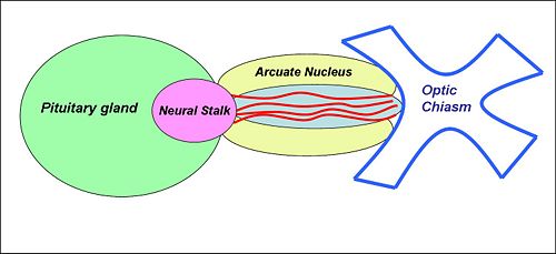
The '''median eminence''' is a specialised region at the base of the brain containing the hypothalamo-hypophysial portal vessels and neuroendocrine nerve endings that release their products into them. The median eminence is a midline structure, immediately below the third cerebral ventricle, bounded laterally by the [[arcuate nucleus]], posterior to the [[optic chiasm]] and rostral to the neural stalk that connects the [[posterior pituitary gland]] to the [[hypothalamus]]. | The '''median eminence''' is a specialised region at the base of the brain containing the hypothalamo-hypophysial portal vessels and neuroendocrine nerve endings that release their products into them. The median eminence is a midline structure, immediately below the third cerebral ventricle, bounded laterally by the [[arcuate nucleus]], posterior to the [[optic chiasm]] and rostral to the neural stalk that connects the [[posterior pituitary gland]] to the [[hypothalamus]]. | ||
{{Image|Median eminence.jpg|right|500px|}} | {{Image|Median eminence.jpg|right|500px|Schematic view of the ventral surface of the brain below the hypothalamus. The median eminence is between the pituitary gland and the optic chiasm, and contains the blood vessels of the hypothalamo-portal system.}} | ||
The median eminence has two zones, an internal zone and an external zone. The internal zone contains the axons of magnocellular neurosecretory neurons of the [[supraoptic nucleus]] and [[paraventricular nucleus]] that project to the posterior pituitary. The external zone contains the convoluted blood vessels of the hypothalamo-pituitary portal system, and the nerve endings of many neuroendocrine neurones. These portal vessels transport releasing factors released by neuroendocrine neurones of the hypothalamus to the anterior pituitary gland. | The median eminence has two zones, an internal zone and an external zone. The internal zone contains the axons of magnocellular neurosecretory neurons of the [[supraoptic nucleus]] and [[paraventricular nucleus]] that project to the posterior pituitary. The external zone contains the convoluted blood vessels of the hypothalamo-pituitary portal system, and the nerve endings of many neuroendocrine neurones. These portal vessels transport releasing factors released by neuroendocrine neurones of the hypothalamus to the anterior pituitary gland. | ||
| Line 11: | Line 11: | ||
* [[corticotrophin releasing hormone]] (CRH) neurones and parvocellular [[vasopressin]] neurones from the [[paraventricular nucleus]] that regulate [[adrenocorticotrophic hormone]] (ACTH) secretion | * [[corticotrophin releasing hormone]] (CRH) neurones and parvocellular [[vasopressin]] neurones from the [[paraventricular nucleus]] that regulate [[adrenocorticotrophic hormone]] (ACTH) secretion | ||
* [[thyrotropic stimulating hormone]] (TRH) neurones that regulate [[thyroid stimulating hormone]] (TSH) secretion. | * [[thyrotropic stimulating hormone]] (TRH) neurones that regulate [[thyroid stimulating hormone]] (TSH) secretion. | ||
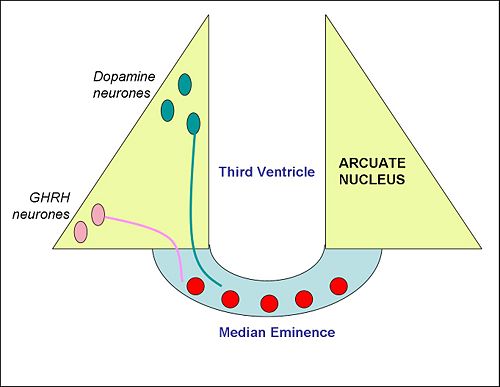
{{Image|Arcuate nucleus.jpg|right|500px|}} | {{Image|Arcuate nucleus.jpg|right|500px| Schematic cross section of the mediobasal hypothalamus, with the median eminence at the bottom.}} | ||
The blood vessels in the external zone of the median eminence are fenestrated, so this zone is outside the [[blood brain barrier]], making it one of the brain's [[circumventricular organs]]. This property means that peptides released from neuroendocrine neurones can freely enter blood vessels. | The blood vessels in the external zone of the median eminence are fenestrated, so this zone is outside the [[blood brain barrier]], making it one of the brain's [[circumventricular organs]]. This property means that peptides released from neuroendocrine neurones can freely enter blood vessels. | ||
Revision as of 10:12, 2 May 2009
The median eminence is a specialised region at the base of the brain containing the hypothalamo-hypophysial portal vessels and neuroendocrine nerve endings that release their products into them. The median eminence is a midline structure, immediately below the third cerebral ventricle, bounded laterally by the arcuate nucleus, posterior to the optic chiasm and rostral to the neural stalk that connects the posterior pituitary gland to the hypothalamus.
The median eminence has two zones, an internal zone and an external zone. The internal zone contains the axons of magnocellular neurosecretory neurons of the supraoptic nucleus and paraventricular nucleus that project to the posterior pituitary. The external zone contains the convoluted blood vessels of the hypothalamo-pituitary portal system, and the nerve endings of many neuroendocrine neurones. These portal vessels transport releasing factors released by neuroendocrine neurones of the hypothalamus to the anterior pituitary gland.
In particular, the median eminence contains neurosecretory nerve endings of
- dopamine neurones from the arcuate nucleus that regulate prolactin secretion
- growth-hormone releasing hormone (GHRH) neurones, also from the arcuate nucleus, that regulate growth hormone secretion
- somatostatin neurones, from the periventricular nucleus, that also regulate growth hormone secretion
- luteinising-hormone releasing hormone (LHRH) neurones from the preoptic area that regulate secretion of both luteinising-hormone (LH) and follicle-stimulating hormone (FSH)
- corticotrophin releasing hormone (CRH) neurones and parvocellular vasopressin neurones from the paraventricular nucleus that regulate adrenocorticotrophic hormone (ACTH) secretion
- thyrotropic stimulating hormone (TRH) neurones that regulate thyroid stimulating hormone (TSH) secretion.
The blood vessels in the external zone of the median eminence are fenestrated, so this zone is outside the blood brain barrier, making it one of the brain's circumventricular organs. This property means that peptides released from neuroendocrine neurones can freely enter blood vessels.
Damage to the median eminence can result in impairment of hormone secretion from the anterior pituitary gland, and in polydipsia (excessive thirst) and polyuria (excessive production of urine) - symptoms of diabetes insipidus as a result of an impairment of vasopressin secretion.

