X-ray: Difference between revisions
imported>Howard C. Berkowitz No edit summary |
mNo edit summary |
||
| (2 intermediate revisions by 2 users not shown) | |||
| Line 1: | Line 1: | ||
{{subpages}} | {{subpages}} | ||
{{TOC|right}} | {{TOC|right}} | ||
'''X-rays''' (aka Röntgen rays, after their discoverer [[Wilhelm Conrad Röntgen]]) are an [[ionizing radiation|ionizing]] type of [[electromagnetic radiation]] in the frequency range of 3×10<sup>16</sup>Hz to 3 × 10<sup>19</sup>Hz. They can be divided into the more energetic hard x-rays (3×10<sup>18</sup>Hz to 3 × 10<sup>19</sup>Hz) adjacent to [[gamma ray]]s, and into soft x-rays (3×10<sup>16</sup>Hz to 3 × 10<sup>18</sup>Hz), adjacent to [[ultraviolet light]]. | '''X-rays''' (aka Röntgen rays, after their discoverer [[Wilhelm Conrad Röntgen]]) are an [[ionizing radiation|ionizing]] type of [[electromagnetic radiation]] in the frequency range of 3×10<sup>16</sup> Hz to 3 × 10<sup>19</sup> Hz. They can be divided into the more energetic hard x-rays (3×10<sup>18</sup> Hz to 3 × 10<sup>19</sup> Hz) adjacent to [[gamma ray]]s, and into soft x-rays (3×10<sup>16</sup> Hz to 3 × 10<sup>18</sup> Hz), adjacent to [[ultraviolet light]]. | ||
They are widely used for structural investigations in all parts of [[materials science]], though care has to be exerted for uses on living tissue, since [[ionizing radiation]] can cause cell damage; see [[acute radiation syndrome]]. | They are widely used for structural investigations in all parts of [[materials science]], though care has to be exerted for uses on living tissue, since [[ionizing radiation]] can cause cell damage; see [[acute radiation syndrome]]. | ||
==Generating X-rays== | ==Generating X-rays== | ||
==Principles of X-ray applications== | ==Principles of X-ray applications== | ||
| Line 48: | Line 49: | ||
==References== | ==References== | ||
{{reflist}} | {{reflist}} | ||
[[Category:Flagged for Review]] | |||
[[Category:Suggestion Bot Tag]] | |||
Latest revision as of 12:01, 9 November 2024
X-rays (aka Röntgen rays, after their discoverer Wilhelm Conrad Röntgen) are an ionizing type of electromagnetic radiation in the frequency range of 3×10¹⁶ Hz to 3 × 10¹⁹ Hz. They can be divided into the more energetic hard x-rays (3×10¹⁸ Hz to 3 × 10¹⁹ Hz) adjacent to gamma rays, and into soft x-rays (3×10¹⁶ Hz to 3 × 10¹⁸ Hz), adjacent to ultraviolet light.
They are widely used for structural investigations in all parts of materials science, though care has to be exerted for uses on living tissue, since ionizing radiation can cause cell damage; see acute radiation syndrome.
Generating X-rays
Principles of X-ray applications
Imaging using X-rays
The most common use of X-rays is in diagnostic imaging. These applications have a source of X-rays, possibly a means of focusing the radiation beam, mechanisms for positioning the source and subject in respect to another, and a device for recording the X-ray beam after it passed through the subject.
X-ray imaging depends on the principle that different materials are more or less opaque to X-rays, and thicker sections of the same material will be more opaque than thinner sections. For example, bone is more opaque than soft body tissue, and thick bone is more opaque than thin bone. In other words, different parts of the subject differently attenuate the X-ray beam. Most often, the image is presented in "negative" form, with whiter parts showing more and the darker parts showing less attenuation.
In a few cases, there is no X-ray source in the system (e.g., X-ray astronomy) or the image is constructed not from the differently attenuated X-rays (e.g., X-ray fluorescence spectroscopy).
Non-imaging X-rays used in evaluating materials
X-rays used to affect materials and tissue
Medical Applications

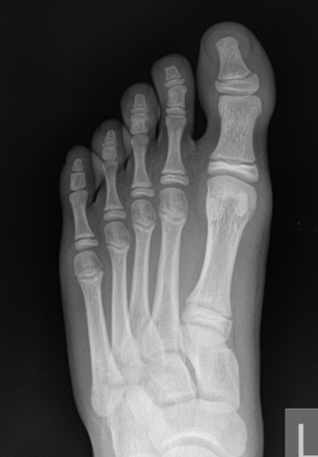
X-ray image of a 10-year old child with Turf Toe, a sprain of the metatarsophalangeal joint of the first toe.
In medicine, the most common use for X-rays is in diagnostic imaging, but there are some applications where the purpose of using the X-rays is to affect tissue, such as radiation oncology.
It is increasingly common to have images read at a distance from the imaging equipment, distance ranging from an office to a continent away. A standard method for transmitting these images is digital imaging and communications in medicine (DICOM).
Medical diagnosis
Practical X-ray machines for diagnostic imaging internal body parts, at a minimum, an #X-ray source, mechanisms both for positioning the X-ray source and the patient, and a means of that records the signals that have gone through the body to form an image.[1]
While they follow the basic principles described here, more advanced medical X-ray techniques, such as X-ray computed tomography, have sufficient differences and refinements that a sub-article is needed for the details.
Medical treatment
X-ray astronomy
This category of astronomy and astrophysics research is one of the few where no X-ray source is needed and the X-ray instrument is purely passive.
X-ray diffraction
- X-ray diffraction [r]: A non-destructive analytical technique which reveal information about the crystallographic structure, chemical composition, and physical properties of materials and thin films, using x-rays. [e]
X-ray fluorescence spectroscopy
X-ray fluorescence spectrosopy is a nonimaging testing methods, applied to nonliving materials, in which the X-rays cause certain atoms in the material to generate, through a special case of the photoelectric effect, energy in a different part of the electromagnetic spectrum than X-rays. [2]
Adverse effects
Radiation dosages for various radiology procedures are available.[3] Radiation from diagnostic imaging may cause cancer.[4][5]
References
- ↑ Digital X-Ray Machine and Camera System, Lattice Semiconductor Corporation
- ↑ X-ray Fluorescence Spectroscopy (XRF), Amptek
- ↑ Mettler FA, Huda W, Yoshizumi TT, Mahesh M (2008). "Effective doses in radiology and diagnostic nuclear medicine: a catalog.". Radiology 248 (1): 254-63. DOI:10.1148/radiol.2481071451. PMID 18566177. Research Blogging.
- ↑ Brenner DJ, Hall EJ (2007). "Computed tomography--an increasing source of radiation exposure.". N Engl J Med 357 (22): 2277-84. DOI:10.1056/NEJMra072149. PMID 18046031. Research Blogging.
- ↑ Einstein AJ, Henzlova MJ, Rajagopalan S (2007). "Estimating risk of cancer associated with radiation exposure from 64-slice computed tomography coronary angiography.". JAMA 298 (3): 317-23. DOI:10.1001/jama.298.3.317. PMID 17635892. Research Blogging.
- Pages using PMID magic links
- CZ Live
- Physics Workgroup
- Chemistry Workgroup
- Health Sciences Workgroup
- Nuclear Engineering Subgroup
- Biophysics Subgroup
- Radiology Subgroup
- Articles written in British English
- All Content
- Physics Content
- Chemistry Content
- Health Sciences Content
- Nuclear Engineering tag
- Biophysics tag
- Radiology tag
- Flagged for Review