File:Brain-MRI-location-guide.png: Difference between revisions
imported>Daniel Mietchen |
imported>Daniel Mietchen |
||
| Line 8: | Line 8: | ||
|pub-country = United States | |pub-country = United States | ||
|notes = | |notes = | ||
|versions = | |versions = [[:Image:CO2-O2-fMRI-A-left.png]] and [[:Image:CO2-O2-fMRI-all.png]] | ||
}} | }} | ||
== Licensing/Copyright status == | == Licensing/Copyright status == | ||
{{CC|by|2.5}} | {{CC|by|2.5}} | ||
Revision as of 17:08, 27 May 2010
Summary
| Title / Description
|
MRI scan of a human brain, with several key anatomical structures labeled. Abbreviations: CC, cerebellum; DM, dorsal midbrain; DMT, dorsal medial thalamus; Hypo, hypothalamus; LAI/RAI, left/right anterior insula; LAP/RAP, left/right posterior insula; LCN/RCN, left/right caudate nucleus; LG, lingual gyrus; LHipp/RHipp, left/right hippocampus; LI/RI, left/right insula; PLT, posterior lateral thalamus; VLT, ventral lateral thalamus. Distances from the anterior commissure and orientation, based on the standard Montreal Neurological Institute space, are (A) 5 mm superior, (B) 5 mm left, (C) 27 mm right, and (D) 19 mm posterior. Distance increases from left-to-right for sagittal (side) views, posterior-to-anterior for coronal views, and inferior to superior for axial (transverse) views. The background image is a high-resolution scan from a single participant (normalized to Montreal Neurological Institute space). |
|---|---|
| Author(s)
|
Macey et al., 2007 |
| Copyright holder
|
Macey et al., 2007 See below for license/re-use information. |
| Source
|
Part of Fig. 1 from Macey PM, Woo MA, Harper RM (2007). "Hyperoxic brain effects are normalized by addition of CO2". PLoS Med 4 (5): e173. DOI:10.1371/journal.pmed.0040173. PMID 17518514. PMC PMC1872042. Research Blogging. [e] |
| Date created
|
June 26, 2006 |
| Country of first publication
|
United States |
| Notes
|
You can edit this page and add notes here which may be useful to people who wish to re-use this media. |
| Other versions
|
Image:CO2-O2-fMRI-A-left.png and Image:CO2-O2-fMRI-all.png |
| Using this image on CZ
|
Copy the code below to add this image to a Citizendium article, changing the size, alignment, and caption as necessary.
|
Please send email to manager A T citizendium.org .
Licensing/Copyright status
This media, Brain-MRI-location-guide.png, is licenced under the Creative Commons Attribution 2.5 Unported License
You are free:
To Share — To copy, distribute and transmit the work; To Remix — To adapt the work.
Under the following conditions:
Attribution — You must attribute the work in the manner specified by the author or licensor (but not in any way that suggests that they endorse you or your use of the work).
For any reuse or distribution, you must make clear to others the licence terms of this work (the best way to do this is with a link to this licence's web page). Any of the above conditions can be waived if you get permission from the copyright holder. Nothing in this licence impairs or restricts the author's moral rights.
Read the full licence.
File history
Click on a date/time to view the file as it appeared at that time.
| Date/Time | Thumbnail | Dimensions | User | Comment | |
|---|---|---|---|---|---|
| current | 19:57, 11 March 2022 | 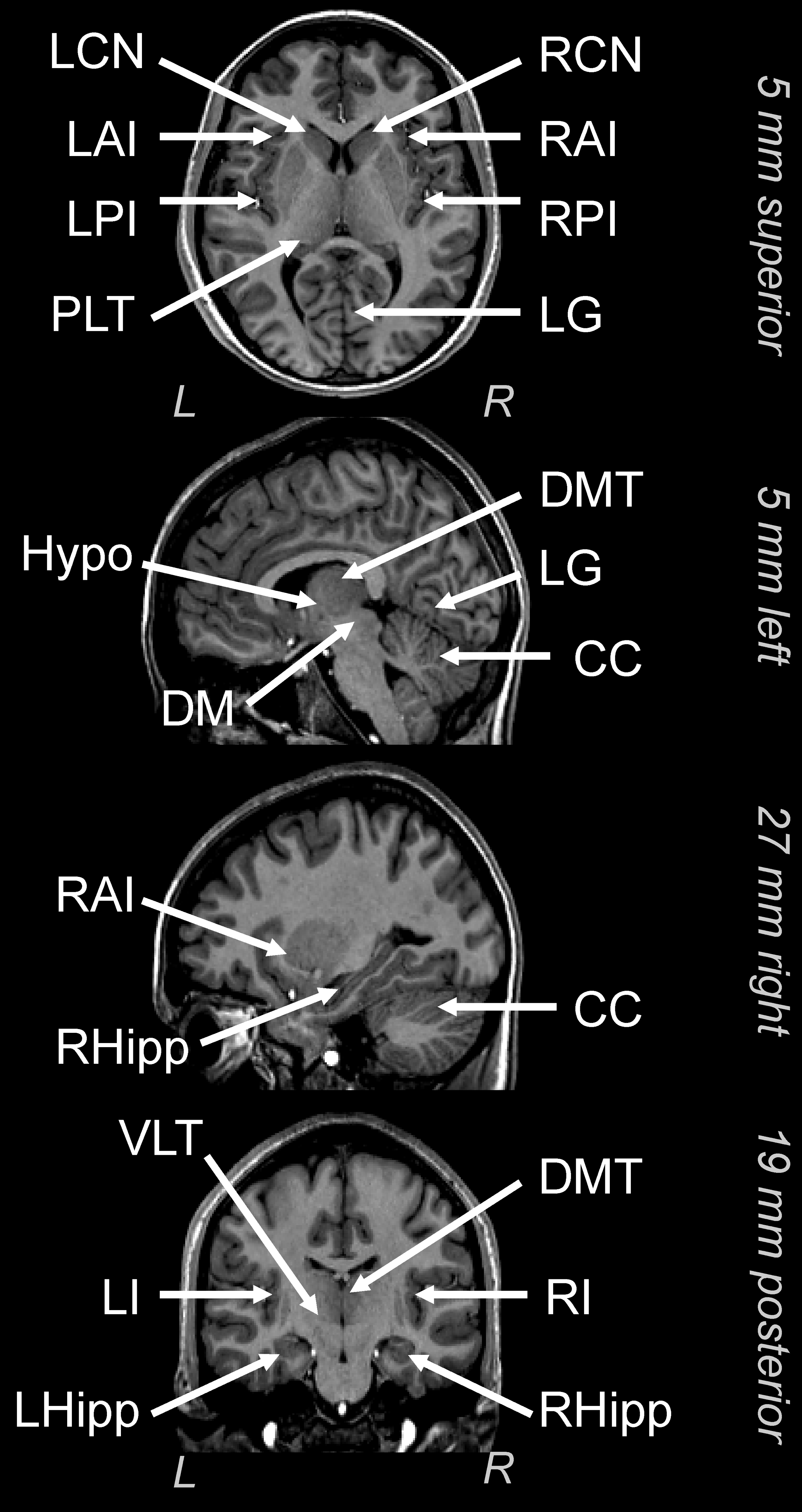 | 2,520 × 4,749 (1.45 MB) | Maintenance script (talk | contribs) | == Summary == Importing file |
You cannot overwrite this file.
File usage
The following page uses this file: